Hydatid Cyst of the Liver: What You Need to Know (And What Others Won't Tell You)
.webp)
Principal Consultant & Unit Head, Liver Transplant & HPB Surgery, Fortis Hospital, Shalimar Bagh, Delhi
Imagine this. You wake up one morning feeling perfectly fine. You have no pain, no yellow skin, no warning signs at all. You go in for a routine ultrasound, maybe for something unrelated like acidity or vague tummy discomfort, and the radiologist finds something unexpected. A fluid-filled cyst. In your liver. When you ask, “How long has this been there?”, the honest answer is: no one knows. It could be five years. It could be fifteen.
This is exactly how a 48‑year‑old businessman from Delhi, Rajesh, first came to see me. He felt healthy, worked long hours, and had never imagined a parasite might be living quietly inside his liver. What makes Rajesh’s story important is not that he felt sick. It is that most people with hydatid cysts feel perfectly fine until something goes terribly wrong. A single ruptured cyst can trigger a life‑threatening allergic reaction, spread parasites throughout the abdomen, or even break through the diaphragm into the lungs.
In this guide, written from my experience as a liver surgeon in Delhi, you will learn what hydatid cysts really are, why they are dangerous even when you feel well, and how we treat them safely and effectively. You will also see some things most websites never mention, such as the risk of anaphylaxis, secondary spread inside the abdomen, and why the exact look of your cyst on scan decides your treatment, not just your symptoms.
Hydatid cyst of the liver is a parasitic infection caused by a tapeworm that grows slowly inside your liver, often without any symptoms, until it ruptures or compresses nearby structures.
The Parasite You Never Knew You Harbored
Here is what most people do not realize. A hydatid cyst does not just appear. It has a long journey before it reaches your liver.
The problem usually starts with dogs. A tiny tapeworm called Echinococcus granulosus lives in the intestine of infected dogs and wild canines. The worm’s eggs pass out in the dog’s stool and contaminate soil, water, vegetables, and animal feed. When a person touches this contamination or eats food washed with unsafe water and then touches their mouth, the eggs enter the body. There is no dramatic moment. No obvious infection. Just a normal day.
Inside the intestine, the egg shell breaks and releases a tiny larva (oncosphere). This larva crosses the intestinal wall, enters the bloodstream, and is carried first to the liver, which acts like a biological filter. In about 70 to 75 percent of human infections, the parasite gets trapped in the liver and slowly develops into a fluid‑filled cyst.
Over months and years, the cyst builds a complex structure:
- An inner germinal layer, which produces baby parasites and tiny particles called “hydatid sand”
- A laminated outer layer, which protects the parasite from your immune system
- A host capsule, formed by your own inflammatory reaction, called the pericyst
The cyst may also develop “daughter cysts” inside it, like small bubbles within a big one. All of this usually happens silently. That is why many patients in India and other endemic regions only discover the cyst during an ultrasound for some unrelated reason.
Why Your Symptoms (or No Symptoms) Can Mislead You
The greatest trap with hydatid cysts is the belief that “no symptoms” means “no problem.”
Early on, most patients feel completely normal. Studies show that up to 70 or even 80 percent of liver hydatid cysts are found incidentally during imaging for another issue. When the cyst is small, your liver simply adjusts. This organ is large and has an amazing reserve, so a 3 or 4‑centimeter cyst often causes no pain, no fever, no jaundice.
Symptoms usually appear when the cyst becomes large (often more than 10 centimeters) or starts pressing on important structures. At that stage you may notice:
- A heavy or dull ache in the upper right side of your abdomen
- Discomfort when lying on the right side
- A feeling of fullness or bloating
- Loss of appetite or mild weight loss
If the cyst presses on the bile ducts, you can develop:
- Yellowing of the eyes and skin (jaundice)
- Dark urine and pale stools
- Itching and more intense pain
The real danger comes when a cyst ruptures.
- If it opens into the bile ducts, patients can suddenly develop fever, severe right‑sided pain, and jaundice due to infection and blockage in the bile ducts (cholangitis).
- If it bursts into the abdominal cavity, the fluid containing millions of parasite particles spills over the abdominal lining. Up to about 10 to 12 percent of these patients can develop anaphylaxis, a severe allergic reaction. The person may break out in hives, feel their throat closing, struggle to breathe, and their blood pressure can collapse within minutes. Without prompt emergency care, this can be fatal.
Rupture can also cause secondary hydatidosis. In this scenario, the spilled parasite particles implant on the surface of different organs and peritoneum (inner abdominal lining), leading to multiple new cysts over time. A single cyst problem turns into a multi‑cyst, multi‑organ problem that is much harder to treat.
So if a scan picks up a hydatid cyst even when you feel fine, you should take it seriously and discuss structured treatment, not wait for symptoms to “prove” it is important.
How Hydatid Cysts Are Diagnosed in Real Life
Patients often ask if a blood test alone can rule this disease in or out. The answer is no. The diagnosis usually needs a combination of history, blood tests, and especially imaging.
Blood tests: helpful but not perfect
Most centers use IgG antibody tests such as ELISA for hydatid disease. In liver cysts, these can have good sensitivity (often above 85 percent) and high specificity. However:
- Early infections can be seronegative (test shows negative even when a cyst is present).
- Old, calcified or inactive cysts may also give negative results.
- Once positive, the test can remain positive for years even after successful treatment.
So a positive test supports the diagnosis, but a negative test does not completely rule it out if the imaging looks typical.
Imaging: the real decision maker
Ultrasound is the first and most important step. It is safe, quick, and widely available. An experienced radiologist can:
- See whether the cyst is simple or has daughter cysts
- Look for floating membranes (the “water‑lily sign”)
- Identify “hydatid sand,” which looks like moving echoes inside the fluid
CT scans provide a 3D view and help:
- Measure exact size and location
- Assess relation to major blood vessels and bile ducts
- Detect multiple cysts or spread to lungs or other organs
Sometimes MRI is used to better understand bile duct involvement or tricky locations, but ultrasound and CT are usually enough in most cases.
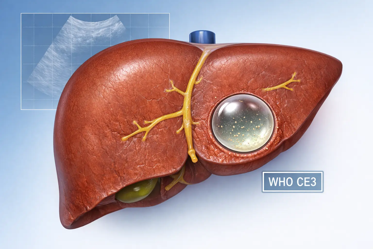
WHO staging: why the appearance on scan decides treatment
Experts use a system called WHO‑IWGE classification, which divides cysts into stages CE1 to CE5 based on how they look on ultrasound. In simple terms:
- CE1, CE2: active, living cysts.
- CE3: transitional, starting to degenerate.
- CE4, CE5: inactive or calcified, often “burnt out.”
This staging tells us if the cyst should be treated with medicine, a needle procedure, surgery, or simply watched over time. In other words, your scan pattern matters more than your symptom level when we plan the safest treatment.
Treatment Options: Medicine, Needles, or Surgery?
There is no single “best” treatment for every hydatid cyst. The right plan depends on the cyst stage, size, location, number of cysts, and your overall health.
Medical treatment (Albendazole)
Albendazole is an anti‑parasitic medicine that can:
- Slow cyst growth
- Help sterilize the cyst before surgery or puncture
- Occasionally shrink small, simple cysts
It is usually given at about 10 to 15 mg per kilogram per day, often for several weeks to months. It is most effective for small, active cysts (like early CE1), and as an addition around procedures rather than as stand‑alone treatment for large or complex ones.
Possible side effects include liver enzyme elevation and effects on blood counts, so regular blood tests are important during long courses.
Percutaneous procedures (PAIR and catheter techniques)
For selected cysts, minimally invasive needle treatments can be effective:
- PAIR (Puncture, Aspiration, Injection, Re‑aspiration) involves placing a needle into the cyst under ultrasound guidance, sucking out fluid, injecting a scolicidal agent such as hypertonic saline or alcohol, and then sucking it out again.
- It works best for simple, single‑chamber cysts without multiple daughter cysts and without clear communication with the bile ducts.
More advanced catheter techniques (like modified catheter drainage) allow repeated washes and removal of solid material from complex cysts.
These methods can reduce hospital stay and avoid large incisions, but they still carry some risk of leakage and rare anaphylaxis, so they must be done in experienced centers with full emergency support.
Surgery: conservative vs radical
Surgery is still the mainstay for many liver hydatid cysts, especially large, complex, or complicated ones.
Broadly, there are two approaches:
In both cases, careful steps are taken during surgery to avoid spilling cyst content into the abdomen. Patients usually receive albendazole before and after the operation to reduce risk of recurrence and secondary spread.
In my practice as a liver transplant surgeon in Delhi, the choice between conservative and radical surgery depends on:
- How much normal liver can be safely preserved
- Whether there are multiple cysts
- How close the cyst is to major vessels or bile ducts
- The patient’s age and other medical conditions
The goal is always the same: remove disease as completely and safely as possible while preserving liver function and quality of life.
Complications You Really Need to Know About
Many basic articles stop at “it can rupture.” Patients deserve a clearer picture.
Anaphylaxis: the hidden emergency
When hydatid fluid suddenly floods the abdominal cavity or bloodstream, your immune system can react violently. This is anaphylaxis, a medical emergency that can cause:
- Sudden itching and rashes
- Swelling of lips, tongue, or throat
- Difficulty breathing or wheezing
- Drop in blood pressure, dizziness, or collapse
This risk is especially important if a cyst ruptures spontaneously or during an accident. It is also a monitored risk during needle‑based treatments and surgery, which is why these procedures are done with full resuscitation facilities.
Secondary hydatidosis
If cyst contents spill and viable parasite elements seed the peritoneum, multiple new cysts can grow across the abdominal cavity in the months that follow. This condition is much harder to treat and often needs several surgeries plus prolonged medical therapy. Surgeons therefore take great care to isolate the operative area and use special solutions to neutralize any spillage inside the abdomen.
Bile duct and lung involvement
Cysts that open into bile ducts can cause repeated episodes of jaundice and infection, and may eventually require a combination of endoscopic procedures and surgery.
Cysts sitting high in the liver, near the diaphragm, can erode into the chest. These can lead to cough, chest infections, and in some cases coughing up cyst material. Such cases often need combined chest and liver surgery in specialized centers.
Life After Treatment: Surveillance and Peace of Mind
Treatment does not end when the operation or procedure is over. Hydatid disease needs follow‑up.
Most patients will be advised to:
- Undergo periodic ultrasound scans, especially during the first 3 to 5 years
- Check liver function and sometimes antibody levels
- Report any new abdominal pain, jaundice, or unexplained fever early
Conservative surgery has higher recurrence rates than radical resection, but even with the best approach there is a small risk that new cysts can appear, either from unnoticed spillage or fresh infection. Regular follow‑up allows us to catch and treat any recurrence early, before it becomes complicated.
Emotionally, many patients feel anxious before each follow‑up scan. That is normal. In my experience, clear communication about the plan, realistic risk numbers, and easy access to the care team help patients regain confidence and return to a normal life.
Can You Prevent Hydatid Disease?
In many ways, yes. Hydatid disease is strongly linked to how animals, meat, and dogs are handled in a community. Places that implemented dog deworming programs and safe slaughter practices have dramatically reduced human cases.
On a personal level, you can reduce your risk by:
- Washing hands thoroughly after contact with dogs or soil
- Avoiding contact with stray dogs as far as possible
- Washing vegetables and fruits well, especially if grown near open fields
- Drinking safe, clean water
- Cooking meat, particularly organ meats, completely before eating
For families in rural or semi‑urban areas with livestock, regular deworming of dogs and proper disposal of infected animal organs are very important to break the cycle of infection.
When Should You See a Liver Specialist?
If your ultrasound or CT report mentions any of the following, you should speak to a liver specialist or hepatobiliary surgeon:
- “Hydatid cyst” or “cystic echinococcosis” of the liver
- Complex cyst with daughter cysts
- Cyst with floating membranes
- Cyst with suspected communication with bile ducts
As a liver transplant surgeon in Delhi, I advise patients not to panic, but not to ignore such findings either. A structured evaluation, correct staging, and a tailored treatment plan are what prevent emergencies and long‑term complications. In selected complex cases, especially when large parts of the liver are involved, the evaluation and skills used in liver transplant practice can be important even if a transplant is not needed.
Conclusion: You Are Not Alone in This
Hearing that you have a “hydatid cyst” in your liver can be frightening. It is natural to think of parasites, rupture, anaphylaxis, or major surgery. But the reality is that with modern imaging, clear staging, and the right combination of medicine, procedures, or surgery, most patients do very well.
If you or a loved one has been diagnosed with a liver hydatid cyst, you do not need to face it alone. A focused discussion with an experienced liver surgeon can clarify:
- What stage your cyst is in
- Which treatment options make sense for you
- What risks are realistic, and how we minimize them
- How your follow‑up will look over the next few years
Are you ready to take the next step and turn a frightening report into a clear, manageable plan?